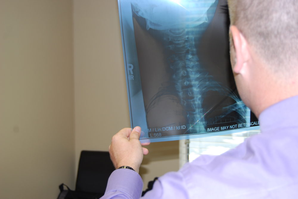
If you have been involved in an auto accident, regardless of the perceived severity, your body has been exposed to a substantial amount of force. The human body is tough, and many times individuals can walk away from an auto accident feeling relatively normal. Modern technology and car crash safety advances are the best they have ever been, and this modern technology is saving lives. Regardless of these advances, every patient that has been involved in an auto accident, needs to complete at least one diagnostic test before deciding if medical treatment is needed or not. This is not hyperbole, every single individual should be evaluated with one fairly inexpensive test. The most important test that every person should have: x-ray.
Auto Accident Basics
Obviously, there is a wide spectrum of auto accidents. Some minor fender benders may only barely scratch the bumper, where as other car wrecks may leave the vehicle nearly unrecognizable. Similarly, injuries that individuals experience follow a similar spectrum of variety. Some patients do not experience any symptoms at all, whereas others may experience permanent disability or even death as a result of the accident. Independent of the perceived severity of the auto accident, the forces of impact between the vehicles involved in the accident have the potential to be transmitted into the people involved in the accident. The risk of soft tissue injury high due to these forces affecting the body in a crash.
Neck Pain and Injury After a Car Crash
Some researchers have estimated that as many as 86 percent of all neck injuries seen clinically resulted from auto accidents, and 85 % of those were from rear-end crashes. These are staggering numbers. The mechanism of neck injury comes from the quick and sudden hyper-extension of the neck with the crash happens, followed by the sudden hyper-flexion of the neck as the head rebounds off the headrest.

Though most individuals do not realize the potential for injury, low-speed accidents can subject car occupants to sudden G-force acceleration too. As much as 2 g on the vehicle and 5 g on the occupant’s head, even if the impact occurred at under 10 mph. At it’s peak, the head’s acceleration can be more than 2.5 times as fast as the peak acceleration of the vehicle that was hit, resulting in significantly more damage to the occupants than to the vehicles. Some researches found head acceleration as high as 11.4 g, even in low-speed collisions.
Diagnostic Testing for the Neck After an Accident
Following a car wreck, we strongly encourage every patient to get a series of diagnostic x-rays of the neck, as well as any spinal areas that hurt. Occasionally, additional x-rays of non-spinal joints may be recommended depending on the physical exam findings.
In a typical, non-fatal auto accident, where the individuals involved are wearing their seat belts, and the airbags either deploy, or do not, the neck is the most susceptible area to injury. This is a result of not having a head or neck restraint, and relying solely on the shoulder/chest and lap belt restraint. Therefor, the head quickly causes the neck to experience soft tissue, whiplash, like injuries through the quick acceleration/deceleration effects of the accident.
An x-ray of the area, regardless of perceived symptoms, is used to rule out two very important diagnoses.
- Acute traumatic spinal fracture
- Acute traumatic ligament tear
Acute Traumatic Spinal Fracture Following a Car Wreck
If you have ever seen a movie where the person involved in a car accident is placed on a long backboard that straps the patient down in order to immobilize them, you have seen a glimpse to how important it is to prevent spinal fractures from creating additional injury. Spinal fractures are a big deal, but they are relatively uncommon compared to soft tissue injuries. Due to their severity though, it is the most common reason for which doctors, especially emergency rooms, choose to x-ray auto accident victims.
Following an accident, if you go to an emergency room, or urgent care facility, whether on your own, or via EMS transport, odds are high that you will have an x-ray of the area that hurts. These initial x-rays are used to rule out spinal fracture as the cause of the pain, and for the most part do a really good job of giving the emergency room physician a picture of what the bones in your spine look like, and if they are fractured or not. As mentioned previously, the majority of these x-rays are negative, and the patient is generally given the diagnosis of spinal pain, though sometimes the diagnosis may include “strain/sprain” of the area involved. The treatment recommendations are usually to take muscle relaxers, and pain reliever medications, and to follow up with your primary care doctor if it doesn’t feel better in a few days or weeks.
Obviously, if there is evidence of a spinal fracture, other procedures may be performed such as bracing, surgery, etc.
Acute Traumatic Spinal Ligament Tears After a Car Accident
Even though emergency room physicians are excellent practitioners that are frequently asked to evaluate individuals immediately following a car accident, acute traumatic spinal ligament tears frequently slips though the cracks of evaluation and diagnosis.
A general rule for using imaging to help diagnose problems in the body, is that x-ray is for bony or hard tissues, while MRI is for soft tissues. Therefore, if a physician needs to be able to diagnose a soft tissue injury such as a disc herniation, or a torn ligament or torn tendon, an MRI is the diagnostic test of choice.
The exception is an acute traumatic spinal ligament injury, which can be identified on a simple x-ray, if the x-ray is performed correctly.
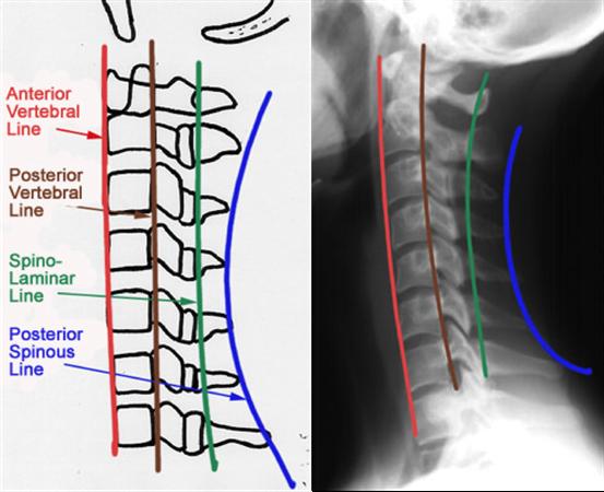
A standard x-ray evaluation of the neck will attempt to determine any abnormal deviations in curvature or posture though analysis of certain points. This evaluation is performed with the patient’s neck in a “neutral” position, which means the neck is not twisted, flexed, or extended. This is a great position to evaluate for fracture, but not a good position for soft tissue involvement. Once fracture is ruled out, other x-ray tests should be performed.
A “Davis Series” of X-Ray Should Follow All Auto Accidents
Clinicians and radiologists have created specific evaluations that can be used in specific situations. According to the International Chiropractic Association, following the trauma of an auto accident, a Davis Series of x-rays should be completed in order to identify soft tissue involvement.
A Davis Series includes the following x-rays:
- A-P cervical
- Lateral cervical
- A-P open mouth (APOM)
- Flexion
- Extension
- Left oblique
- Right oblique
The A-P cervical (front to back), and lateral cervical (side view), are routine x-rays, and are nearly always evaluated in order to rule out fracture. Occasionally, the A-P open mouth (APOM) view will be ordered in an emergency room setting, though it is it more common in chiropractic offices. This x-ray prioritizes viewing the top two bones in the neck (C1/C2 or Atlas/Axis).
The Davis Series includes four additional x-rays, the flexion/extension views and the left/right oblique views. The oblique views can show fractures that may not have been identified with the standard series, but also have the ability to show an opening between the joints called the “neural foramina.” The neural foramina is a small opening where the nerves leave the spinal cord, and go to muscles, organs, and other tissues in the body. Disruption of the neural foramina can lead to radiating symptoms, numbness, tingling, and more.
The flexion/extension x-rays are the most important x-rays for determining soft tissue, ligament, injury. As described earlier, certain “lines” can be drawn over an x-ray in order to ensure normal alignment. These lines should be maintained when a person fully flexes or extends the spine. A disruption in one of these lines is a big deal, yet these tests are often ignored.
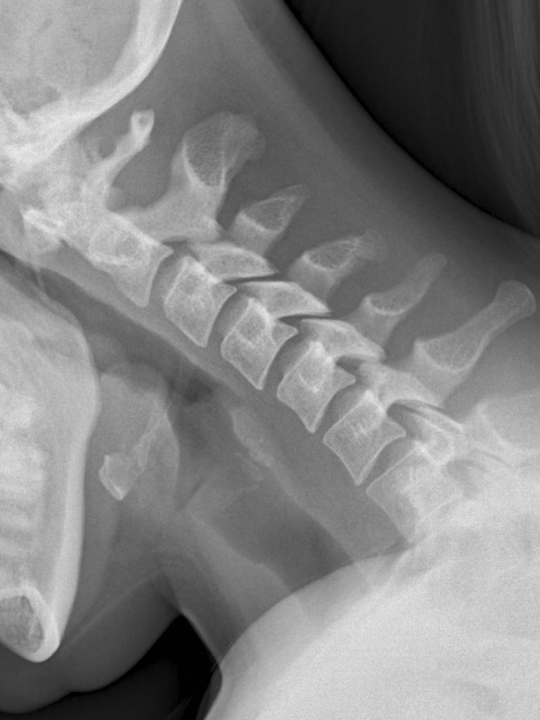
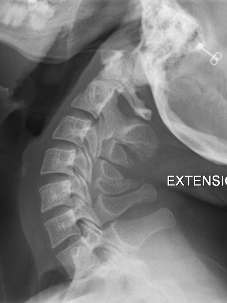
These images above demonstrate normal flexion and extension neck x-rays, and how the various lines are maintained. These images do not demonstrate a tear in the soft tissue ligaments that support and stabilize the neck.
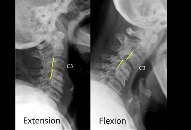
Image courtesy of Lennard A. Nadalo, MD.
Alternatively, in this example from Medscape, an elderly patient experienced neck pain with movement (non-trauma induced in this situation). Normal alignment is seen on the extension view (left), but on the flexion view (right), there is disruption of the middle column, with movement of C3 anterior to C4 (yellow lines). This is consistent with a ligamentous disease. Chronic degenerative changes of the facet joints and the supporting spinal ligaments may result in moderate cervical spinal instability, as in this case. In addition to chronic degeneration, acute traumatic injury can also create this finding on x-ray.
Have A Complete X-Ray Evaluation After an Auto Accident
Due to the frequency of soft tissue injuries that accompany a car wreck, we perform x-ray evaluations on every patient that comes to our office with accident related injuries. It is essential that we identify all of the tissues involved with the accident so that the correct diagnosis, and treatment can be performed. Unfortunately, many injuries go diagnosed and untreated because patients feel “fine” after the accident, or upon leaving the emergency room the patient is told that there are no broken bones so they assume that everything else is OK as well.
If you have recently been involved in an auto accident, even if you do not come to our office for treatment, we encourage you to advocate for complete x-ray testing in order to determine the extent of your injuries. If we can be of assistance in helping in your recovery, please contact our office and schedule with one of our doctors.
You may also like this

I was Involved In a Car Accident, Now What?!

5 Mistakes to Avoid with Insurance Companies After a Car Wreck